Fascinating talk by Dale Moulding.
In this series of blogs we share the webinars organised by ConCode. Conserv is a sponsor of ConCode and our very own Melissa King is one of its founding members. Find more about our partnerships here.
What is ConCode?
ConCode is a community of coders that work in the cultural heritage field and find each other on the ConCode Slack Channel. If this is something that interests you, you can join the Slack Channel here.
ConCode is committed to highlighting the role of coding and development in the world of cultural heritage management. This is why they run webinars and events on various related topics. This latest talk focuses on how to use the open source Fiji software to batch run image analyses for archaeometry and archaeology research.
What can you do with Fiji software?
Dale Moulding, light microscope specialist working at UCL in London, talks about his experience working on image analysis applied to the fields of archaeometry and cultural heritage.
Webinar breakdown
- Introduction to the Fiji / ImageJ software.
- Description of what digital images are and insights into basic image processing steps.
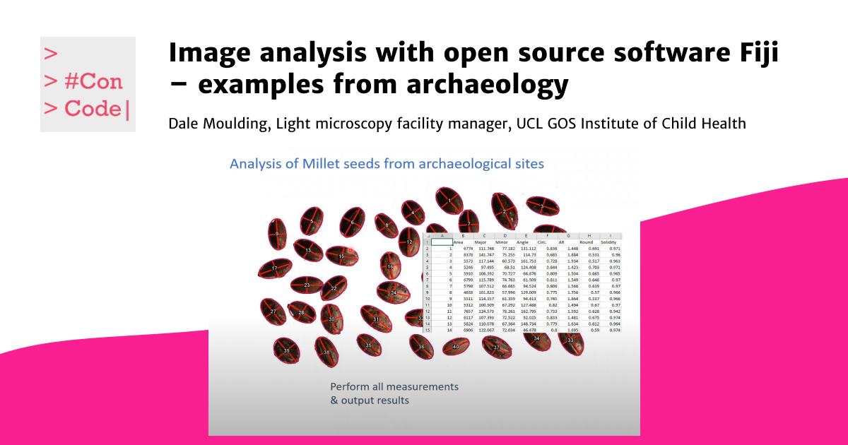
- Case study 1: The application of image analysis to automate fossilised Millet seed measurements.
- Case study 2: Fiji software as a tool to measure potsherds morphometrics for post-depositional erosion and fragmentation.
Dale will show how image analysis using a tool like Fiji helped him unravel some mysteries about the history of the archaeological artefacts investigated and the life of the people who used them.
Watch the recording of the lecture here:
Who is Dale Moulding?
“My name is Dale Moulding and I am a light microscope specialist at the GOS Institute of Child Health in London (UK). My background (16 years research) is in immunology and cell biology, with a heavy emphasis to applying microscopy to understand immune processes. In the last 5 years, I have been running the confocal and light microscopy core facility at my institute where I train users in the most appropriate imaging system for their research and provide custom image analysis protocols. As part of this, I run an open access image analysis course through which I have helped analyse mostly biological samples, but also ice-cream, seaweed, paintings, Persian rugs & archaeological samples.”
To know get more information about the facility and the course, follow the links below:
https://www.ucl.ac.uk/ich/core-scientific-facilities-centres/confocal-microscopy(facility)